
Some 113,000 patients are currently on an organ waiting list, unsure whether they will receive the life-saving treatment that they desperately need. Artificially grown, or 3D-printed organs are viewed as a solution to the organ shortage, but to date scientists have been unable to create living tissue with the cellular density and organ-level functions required to produce reliable results.
However, a team of researchers from Harvard’s Wyss Institute for Biologically Inspired Engineering and John A. Paulson School of Engineering and Applied Sciences claim to have created a new technique that overcomes these issues.
Go deeper with GlobalData


Access deeper industry intelligence
Experience unmatched clarity with a single platform that combines unique data, AI, and human expertise.
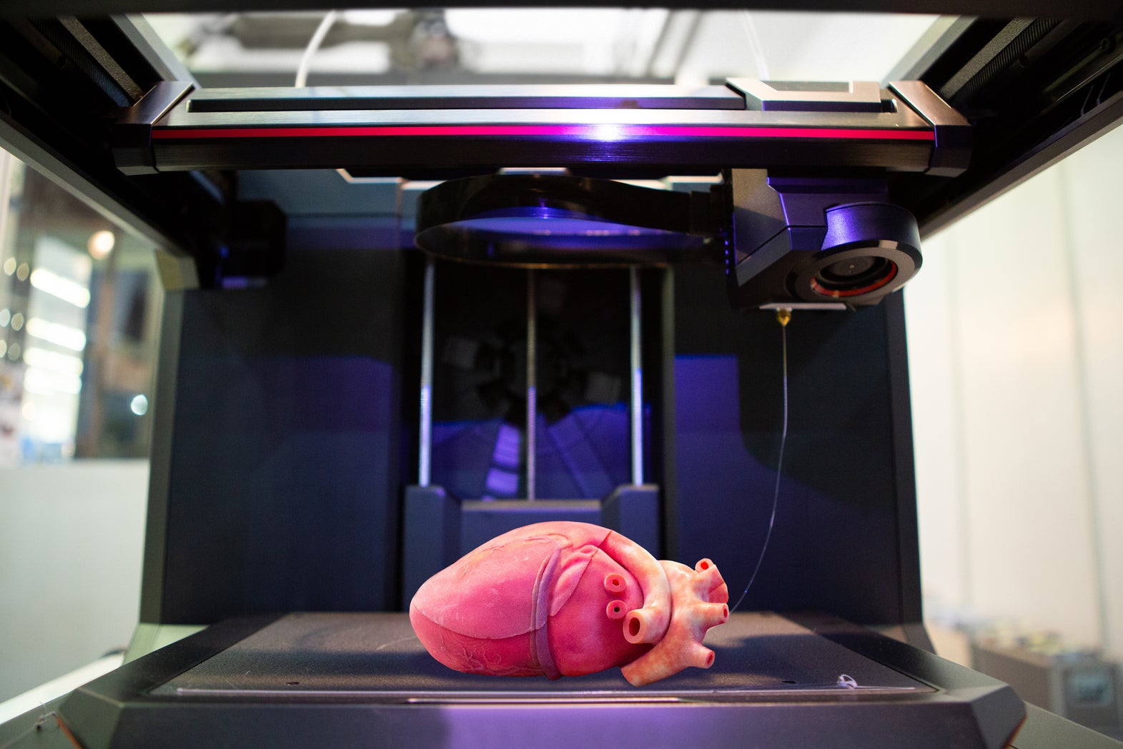
3D printing organs: The SWIFT technique
The Sacrificial writing into functional tissue (SWIFT) technique involves forming hundreds of thousands of stem-cell derived aggregates into organ building blocks (OBBs), using a mixture of stem cells and a specially-made extracellular solution to create a living network containing approximately 200 million cells per millilitre.
This mixture is almost liquid at cold temperatures, allowing it to be shaped without causing damage to the cells. However, the mixture solidifies when heated, making it an ideal material to create 3D-printed organs with.
A vascular network is then embedded within it, which allows oxygen and nutrients to reach the cells, mimicking the blood vessels found inside real organs.
Tests showed that tissues with these embedded vascular channels remained stable, while tissue grown without them began to experience cell death within 12 hours.
To further test the technique’s potential, the team printed a network of heart-derived cells and flowed media through its vascular network for more than a week. Results show that the OBBs fused together over time, forming more solid tissue and growing in strength by more than 2,000%, mimicking key features of the human heart.
“Forming a dense matrix from these OBBs kills two birds with one stone: not only does it achieve a high cellular density akin to that of human organs, but the matrix’s viscosity also enables printing of a pervasive network of perfusable channels within it to mimic the blood vessels that support human organs,” Sebastien Uzel, a research associate involved in the study, explained.
“This is an entirely new paradigm for tissue fabrication,” researcher Mark Skylar-Scott said. “Rather than trying to 3D-print an entire organ’s worth of cells, SWIFT focuses on only printing the vessels necessary to support a living tissue construct that contains large quantities of OBBs, which may ultimately be used therapeutically to repair and replace human organs with lab-grown versions containing patients’ own cells.”
Further testing is currently being tested out by researchers from the Wyss Institute, Boston University and the Massachusetts Institute of Technology.
The research has been published in the Science Advances journal.
Read more: How close are 3D printed organs to reality?